
Visceral Fat: What It Is and How to Measure It

Visceral fat accumulates around organs and drives metabolic disease. Learn how to measure it, what levels matter, and how to reduce it.
This page is for general information and does not constitute medical advice, diagnosis, or treatment. Results vary between individuals. GLP-1 medication (semaglutide — Ozempic, Wegovy; tirzepatide — Mounjaro) is a prescription medicine, dispensed and administered only after assessment by a licensed physician. This page is medical information, not an advertisement or an offer of treatment. Always consult a qualified physician about your condition. See our full medical disclaimer.
Not all fat sits in the same place or carries the same risk. The fat you can pinch sits beneath the skin. The fat you cannot see wraps around your liver, intestines, and kidneys. That second type is visceral fat, and it is the one that matters most for metabolic health. Two people with identical body weights and waist measurements can have very different visceral fat levels. One may carry most of their adipose tissue subcutaneously. The other may store it deep in the abdomen, around the organs. The second person faces higher risk of insulin resistance, cardiovascular disease, and systemic inflammation, even if they appear lean.
What Visceral Fat Is and Where It Sits
Visceral fat is adipose tissue that accumulates in the abdominal cavity, surrounding the liver, pancreas, intestines, and other visceral organs. Unlike subcutaneous fat, which sits just beneath the skin and provides insulation and energy storage, visceral fat is metabolically active and secretes inflammatory cytokines and hormones that disrupt normal physiology.
The body stores fat in distinct compartments. Subcutaneous adipose tissue accounts for most total body fat in healthy individuals. Visceral adipose tissue typically represents 10 to 20 percent of total fat in men and 5 to 8 percent in women, though this varies widely by age, genetics, and lifestyle. Small increases in visceral fat carry disproportionate metabolic consequences.
Key differences between visceral and subcutaneous fat:
- Location: Visceral fat sits inside the peritoneal cavity; subcutaneous fat lies between skin and muscle.
- Metabolic activity: Visceral adipocytes release more free fatty acids directly into the portal circulation, affecting liver metabolism.
- Inflammation: Visceral fat produces higher levels of interleukin-6, tumor necrosis factor-alpha, and other pro-inflammatory markers.
- Insulin sensitivity: Even modest visceral fat accumulation impairs glucose uptake and insulin signaling.
Visceral fat accumulation increases with age, particularly after menopause in women. Genetics play a role, but diet, physical activity, sleep quality, and chronic stress explain most of the variation between individuals.

Why Visceral Fat Drives Metabolic Disease
The location of visceral fat explains its metabolic impact. Adipocytes in the visceral depot drain directly into the hepatic portal vein, delivering free fatty acids and adipokines straight to the liver. This constant influx promotes hepatic insulin resistance, triglyceride synthesis, and ectopic fat deposition.
Visceral adipose tissue pathophysiology involves chronic low-grade inflammation. Visceral adipocytes become hypertrophic and dysfunctional, secreting inflammatory cytokines that attract macrophages and other immune cells. The resulting inflammation spreads systemically, affecting insulin signaling in muscle and liver.
Consequences of Elevated Visceral Fat
| Organ System | Effect |
|---|---|
| Liver | Increased triglyceride synthesis, non-alcoholic fatty liver disease, impaired glucose regulation |
| Pancreas | Beta-cell dysfunction, reduced insulin secretion, elevated fasting glucose |
| Cardiovascular | Endothelial dysfunction, atherosclerosis, hypertension, increased cardiac workload |
| Systemic | Chronic inflammation, oxidative stress, insulin resistance across tissues |
Visceral fat also disrupts adipokine balance. Leptin levels rise, but leptin resistance prevents normal satiety signaling. Adiponectin, which enhances insulin sensitivity and has anti-inflammatory properties, declines as visceral fat increases. The ratio of leptin to adiponectin becomes a marker of metabolic dysfunction.
The risk is dose-dependent. Each additional kilogram of visceral adipose tissue measurably increases the likelihood of type 2 diabetes, cardiovascular events, and all-cause mortality. The relationship holds even in individuals with normal body mass index, a phenomenon sometimes called metabolically obese normal weight.
How to Measure Visceral Fat
Body weight and BMI do not differentiate between subcutaneous and visceral fat. Waist circumference offers a rough proxy. Men with waist circumference above 102 centimeters and women above 88 centimeters likely have elevated visceral fat, but the measurement cannot distinguish between fat compartments or account for muscle mass and skeletal frame.
Direct measurement requires imaging:
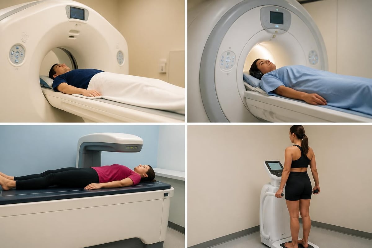
- Computed tomography (CT): A single slice at the L4-L5 vertebral level quantifies visceral and subcutaneous adipose tissue area with high precision. Radiation exposure limits repeated use.
- Magnetic resonance imaging (MRI): Multi-slice abdominal MRI provides volumetric visceral fat assessment without radiation. Gold standard for research, though costly and time-intensive.
- Dual-energy X-ray absorptiometry (DXA): Estimates visceral adipose tissue using algorithms based on android fat distribution. Less accurate than CT or MRI but widely available and low-radiation.
- Bioelectrical impedance (BIA): Some devices estimate visceral fat using proprietary algorithms. Accuracy varies by device and hydration status; useful for tracking trends, not absolute values.
CT and MRI remain the reference standards. A single CT slice at L4-L5 correlates strongly with total visceral adipose tissue volume. Clinically, a visceral fat area above 100 square centimeters on CT signals elevated metabolic risk. Below 100 square centimeters is generally considered lower risk, though individual thresholds vary by ethnicity and sex.
At Healthi Life, visceral fat quantification is part of a comprehensive metabolic assessment. A physician reviews imaging alongside biomarkers, lipid panels, glucose regulation, and inflammatory markers to build a complete picture. The decision to intervene is never made on imaging alone.
What Levels of Visceral Fat Matter
Reference ranges depend on the imaging modality and the population studied. CT-based thresholds are the most established. Visceral adipose tissue area below 100 square centimeters at L4-L5 is associated with lower metabolic risk in most populations. Areas between 100 and 160 square centimeters indicate moderate risk. Above 160 square centimeters correlates with substantially higher incidence of type 2 diabetes and cardiovascular disease.
Reference Ranges by Sex (CT at L4-L5)
| Category | Men (cm²) | Women (cm²) |
|---|---|---|
| Low risk | < 100 | < 80 |
| Moderate risk | 100–160 | 80–130 |
| High risk | > 160 | > 130 |
Women generally accumulate less visceral fat than men at any given BMI, but post-menopausal redistribution shifts fat storage toward the visceral depot. Ethnicity also modifies risk thresholds. South Asian and East Asian populations show metabolic dysfunction at lower visceral fat levels than European or African populations.
Visceral fat increases with age. A 50-year-old with 110 square centimeters of visceral adipose tissue may face different absolute risk than a 30-year-old with the same measurement, though both are above the low-risk threshold. The physician interprets the number in context: family history, current metabolic markers, physical activity level, and other risk factors.
Tracking change over time matters more than a single measurement. A reduction of 20 to 30 square centimeters visceral fat area, achieved through lifestyle modification or medical intervention, produces measurable improvements in insulin sensitivity and inflammatory markers.
How to Reduce Visceral Fat
Visceral fat responds to caloric restriction more readily than subcutaneous fat. A sustained energy deficit, even a modest one, preferentially mobilizes visceral adipose tissue. Weight loss of 5 to 10 percent of body weight typically reduces visceral fat by 20 to 30 percent, with corresponding improvements in glucose metabolism and lipid profiles.
Exercise and Visceral Fat
Aerobic exercise reduces visceral fat independent of weight loss. A recent network meta-analysis found that moderate-intensity continuous training and high-intensity interval training both effectively reduce visceral adipose tissue, with high-intensity protocols showing slightly greater efficiency per unit of time. Resistance training also contributes, particularly when combined with aerobic work.
Effective exercise protocols:
- Moderate-intensity aerobic exercise: 150 to 300 minutes per week
- High-intensity interval training: 75 to 150 minutes per week
- Resistance training: Two to three sessions per week, targeting major muscle groups
- Combined aerobic and resistance programs show additive effects
The dose matters. Weekly exercise volume below 150 minutes shows minimal impact on visceral fat. Above 200 minutes, the dose-response curve flattens, though metabolic benefits continue to accrue. Consistency over months, not intensity over days, drives the change.
Sleep and stress also influence visceral fat accumulation. Chronic sleep restriction raises cortisol and promotes visceral adiposity. Poor sleep quality correlates with higher visceral fat area independent of total sleep duration. Similarly, chronic psychological stress and elevated cortisol favor visceral over subcutaneous fat deposition.

Nutritional Approaches
No single macronutrient composition eliminates visceral fat, but certain patterns show consistent benefit. Low-carbohydrate diets, Mediterranean diets, and time-restricted eating all reduce visceral fat when total energy intake is controlled. The common factor is sustained caloric deficit and avoidance of ultra-processed foods.
Dietary factors that influence visceral fat:
- Refined carbohydrates and added sugars: Promote hepatic lipogenesis and visceral fat accumulation
- Saturated fat intake: High intake, particularly from processed sources, correlates with visceral adiposity
- Fiber: Soluble fiber improves insulin sensitivity and may modestly reduce visceral fat over time
- Protein: Higher protein intake (1.6 to 2.2 grams per kilogram body weight) preserves lean mass during weight loss and may enhance visceral fat reduction
Alcohol contributes to visceral fat, particularly in men. Even moderate intake (more than one drink per day) is associated with increased visceral adipose tissue area. The effect is dose-dependent.
Medical and Pharmacological Interventions
When lifestyle modification alone does not achieve target visceral fat reduction, pharmacological options exist. GLP-1 receptor agonists reduce visceral fat and hepatic fat content beyond what is expected from weight loss alone. Metformin improves insulin sensitivity and may modestly reduce visceral adiposity in individuals with insulin resistance or prediabetes.
Bariatric surgery produces the largest and most rapid reductions in visceral fat. Post-operative visceral adipose tissue area decreases by 40 to 60 percent within the first year. Metabolic improvements, including remission of type 2 diabetes, correlate more strongly with visceral fat loss than with total weight loss.
The decision to use medication or surgery is made after a full review of metabolic markers, imaging, and the individual's response to structured lifestyle intervention. Not everyone with elevated visceral fat requires pharmacotherapy. Some respond fully to exercise and dietary change alone.
Visceral Fat and Long-Term Health Outcomes
Visceral fat predicts mortality independent of BMI. In large cohort studies, individuals in the highest quartile of visceral adipose tissue area have two to three times the risk of cardiovascular death compared to those in the lowest quartile, even after adjusting for total body fat, blood pressure, cholesterol, and smoking status.
The relationship extends beyond cardiovascular disease. Visceral fat is associated with increased risk of certain cancers, including colorectal, breast, and pancreatic cancer. The mechanisms likely involve chronic inflammation, insulin resistance, and altered sex hormone metabolism.
Visceral fat also affects cognitive health. Higher visceral adipose tissue correlates with faster cognitive decline and increased dementia risk in longitudinal studies. The link may be mediated by vascular damage, chronic inflammation, or direct effects of adipokines on brain function.
Reducing visceral fat improves outcomes. Randomized trials show that interventions lowering visceral adipose tissue area by 20 percent or more produce measurable reductions in inflammatory markers, improvements in insulin sensitivity, and better lipid profiles. These changes translate to lower calculated cardiovascular risk scores.
Who Should Measure Visceral Fat
Not everyone needs direct visceral fat measurement. Individuals with normal waist circumference, good metabolic health, and no family history of diabetes or cardiovascular disease may not benefit from imaging. The decision is clinical, made after reviewing symptoms, lab results, and risk factors.
Candidates for visceral fat assessment:
- Individuals with metabolic syndrome or prediabetes, regardless of BMI
- Those with normal BMI but elevated waist circumference or central adiposity
- People with family history of type 2 diabetes or early cardiovascular disease
- Patients with non-alcoholic fatty liver disease or unexplained insulin resistance
- Anyone beginning a structured weight-loss or longevity program who wants baseline data
Visceral fat measurement provides a baseline and a target. It turns abstract metabolic risk into a concrete number. Re-measurement after three to six months of intervention shows whether the protocol is working or needs adjustment.
The imaging itself is straightforward. A CT scan at L4-L5 takes minutes. DXA requires slightly more time but involves lower radiation. MRI is longer but offers the most detail. The physician reviews the result alongside other data to decide next steps.
Visceral Fat in the Context of a Longevity Program
Visceral fat is one marker among many. A longevity program does not start with imaging alone. It begins with a diagnostic baseline: biomarkers, metabolic panels, inflammatory markers, hormone levels, and, when indicated, imaging to quantify visceral and hepatic fat.
The physician uses the full dataset to build a protocol. If visceral fat is elevated, the plan addresses it through structured exercise, nutritional guidance, and, if appropriate, pharmacological support. Re-testing at defined intervals shows whether the intervention is effective.
Visceral fat reduction is not an end in itself. It is a means to improve insulin sensitivity, reduce systemic inflammation, and lower long-term disease risk. The goal is a sustained metabolic shift, not a single number on a scan.
A well-designed program measures, adjusts, and measures again. Visceral fat becomes part of a larger picture: How is glucose regulation changing? Are inflammatory markers declining? Is liver function improving? The physician makes decisions based on the trend across all markers, not visceral fat alone.
Visceral fat is invisible but measurable, and its reduction is one of the most effective metabolic interventions available. The process begins with accurate assessment, continues with a structured protocol, and requires regular re-evaluation. At Healthi Life, every plan follows this sequence: measure the baseline, understand the data, decide on the protocol, and track the change under medical supervision. If you want to know where you stand and what to do next, visit Healthi Life.
This page is for information only and is not medical advice. Medical consultation and prescription are available online and on site at Healthi Life, Ekkamai, Bangkok.